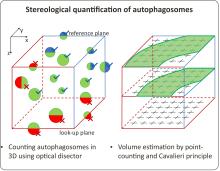
The workflow enables the quantification of autophagosomes in Arabidopsis thaliana root epidermal cells in 3D. It combines immunolabeling of an autophagosome marker ATG8 with commercially available anti-ATG antibody and the subsequent stereological quantification of the immunolabeled particles. The immunolabeled samples are imaged with a confocal microscope and Z-stacks are acquired. The stereological methods involve counting of the immunolabeled autophagosomes using the optical disector and the determination of volume of the examined tissue with the Cavalieri principle and point counting. The image analysis is performed entirely in Fiji and requires no further special software. The method provides a powerful toolkit for unbiased and reproducible quantification of autophagosomes and offers a convenient alternative to the standard of live imaging with FP-ATG8 markers.
Typ publikace
Periodikum nebo kniha
Datum vydání

